Intravascular Imaging
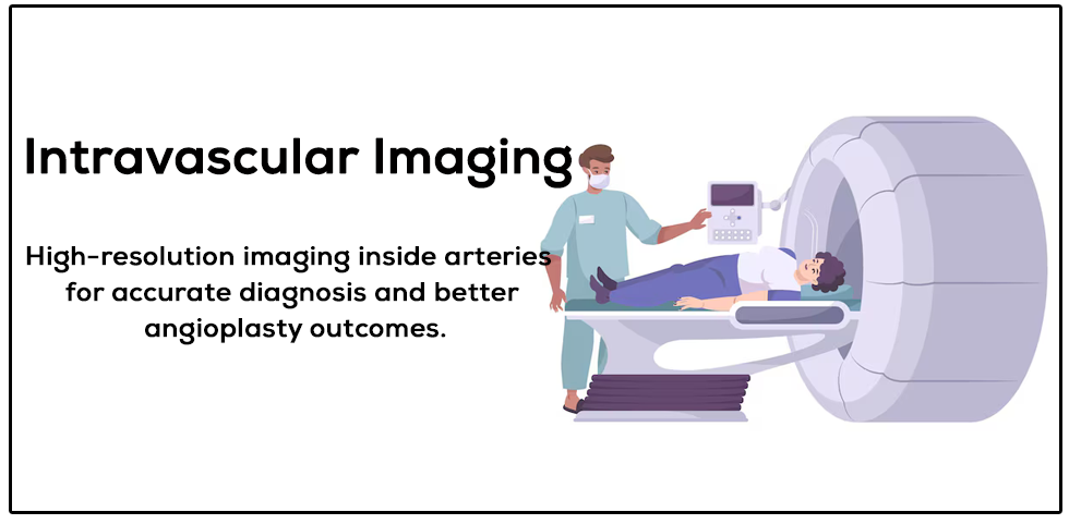
Intravascular Imaging is an advanced diagnostic technique used during coronary procedures to provide detailed, real-time images from inside the blood vessels. It helps cardiologists accurately assess the severity, location, and nature of artery blockages, leading to more precise and effective treatment.
This technology uses specialized tools such as IVUS (Intravascular Ultrasound) and OCT (Optical Coherence Tomography) to visualize the inner structure of the coronary arteries. By offering high-resolution images, intravascular imaging allows better decision-making during procedures like angioplasty and stent placement.
It significantly enhances the safety and success of cardiac interventions by ensuring optimal stent positioning and reducing the risk of complications.
When is Intravascular Imaging Recommended?
- During coronary angioplasty for better accuracy
- To assess complex or unclear artery blockages
- For optimal stent placement and expansion
- In cases of recurrent blockage or stent failure
- High-risk or complicated coronary artery disease
Benefits of Intravascular Imaging
- Provides detailed, high-resolution images of arteries
- Improves accuracy of diagnosis and treatment
- Ensures proper stent placement and expansion
- Reduces risk of complications and repeat procedures
- Enhances overall success of angioplasty
Types of Intravascular Imaging
- IVUS (Intravascular Ultrasound)
- OCT (Optical Coherence Tomography)
Why Choose Dr. Sujeet Narain for Intravascular Imaging?
- Expertise in advanced imaging-guided interventions
- Experience in managing complex coronary cases
- Use of latest IVUS and OCT technologies
- High precision and patient safety standards
- Improved outcomes with personalized treatment approach